Gallery
granulocyte images: illustrations and micrographs. All credits and licenses listed.
Micrographs
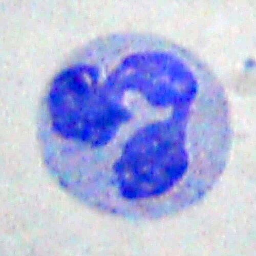
Neutrophil in a peripheral blood smear, Wright stain. The multilobed nucleus and pale cytoplasm are characteristic of the circulating PMN.
Bob Blaylock, Wikimedia Commons · CC BY-SA 3.0 · source
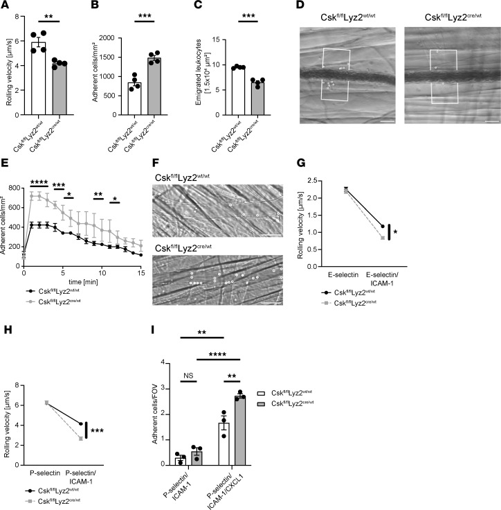
Amini, Schemmelmann, Heming et al 2025, JCI Insight · CC BY 4.0 · source
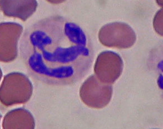
Neutrophil in a peripheral blood smear showing the characteristic segmented nucleus with 3 to 5 lobes.
Dr. Roshan Nasimudeen, Wikimedia Commons · CC BY-SA 3.0 · source
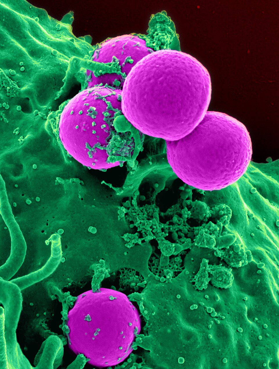
Scanning electron micrograph of a human neutrophil (blue) engulfing MRSA bacteria (yellow). The cell extends pseudopods around the bacteria in a classic phagocytic embrace.
National Institutes of Health (NIH) · Public domain · source
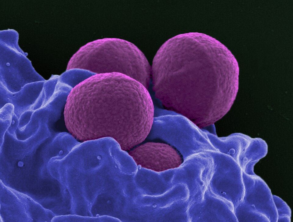
Scanning electron micrograph of a neutrophil (orange-brown) with MRSA bacteria (purple). A second image from the NIH SEM series on neutrophil-pathogen interaction.
National Institutes of Health (NIH) · Public domain · source
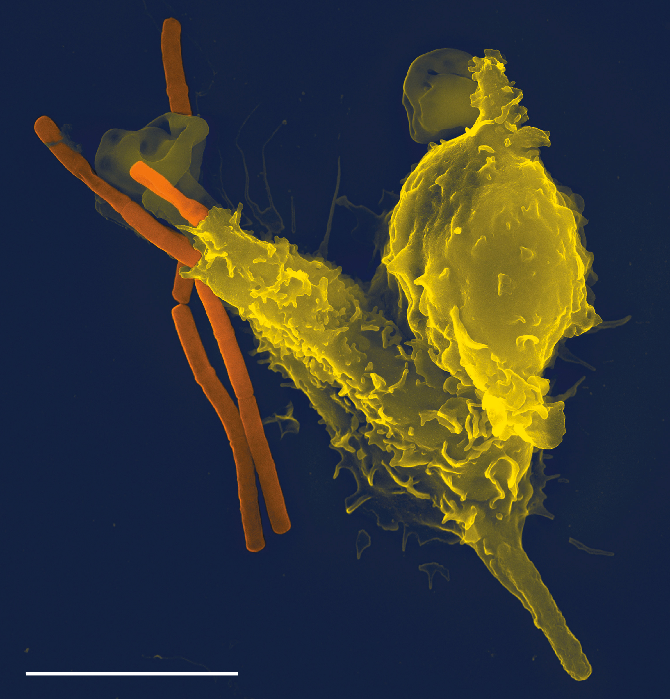
Scanning electron micrograph of a neutrophil (orange) engaging a Bacillus anthracis chain (green). Taken by Volker Brinkmann, the scientist who discovered neutrophil extracellular traps.
Volker Brinkmann, Wikimedia Commons · CC BY 2.5 · source
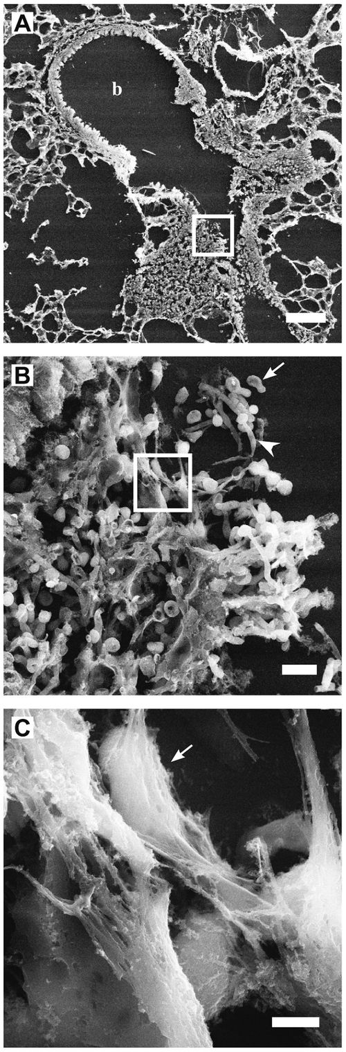
Scanning electron micrograph of neutrophil extracellular traps (NETs): fine fibres of decondensed chromatin with embedded granule proteins enmeshing Staphylococcus aureus.
Urban CF et al. 2009, PLoS Pathogens, Wikimedia Commons · CC BY 2.5 · source
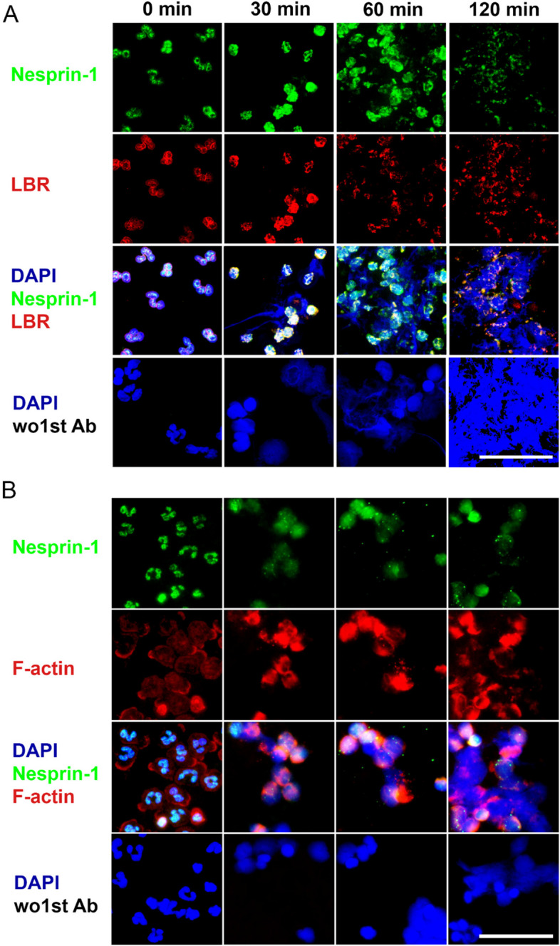
Immunofluorescence of NETs: neutrophils (blue DAPI nuclei) releasing decondensed chromatin co-stained with MPO (red) and citrullinated histone H3 (green).
Singh, Zlatar, Munoz-Becerra et al 2024, Cell Communication and Signaling · CC BY 4.0 · source
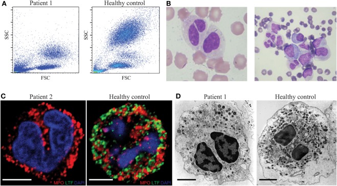
Transmission electron micrograph of a human neutrophil showing the multilobed nucleus and electron-dense azurophilic granules in the cytoplasm.
Serwas, Huemer, Dieckmann et al 2018, Frontiers in Immunology · CC BY · source
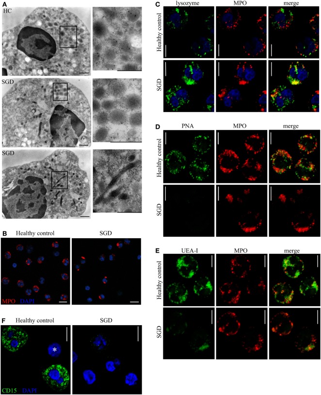
Immunoelectron micrograph of a neutrophil granule showing immunogold labeling of specific granule contents. Granule populations are distinguished by their electron density and marker proteins.
Serwas, Huemer, Dieckmann et al 2018, Frontiers in Immunology · CC BY · source
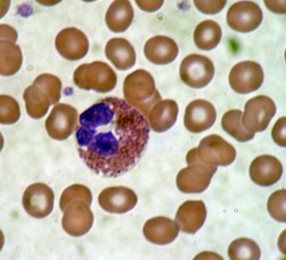
Eosinophil in a peripheral blood smear, Giemsa stain. The bilobed nucleus and large brick-red granules packed with major basic protein are the defining features.
Bobjgalindo, Wikimedia Commons · CC BY-SA 4.0 · source
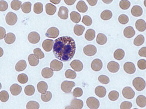
Eosinophil among red blood cells, Giemsa stain. The bright orange-red cytoplasmic granules are visible against the pale erythrocytes.
Dr Graham Beards, Wikimedia Commons · CC BY-SA 3.0 · source
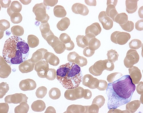
Activated eosinophils in idiopathic hypereosinophilic syndrome. Degranulated cells with vacuolated cytoplasm; some show extracellular granule deposits, a hallmark of cytolytic degranulation.
NIAID, Wikimedia Commons · CC BY 2.0 · source
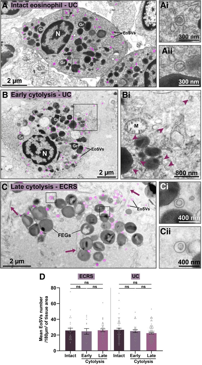
Transmission electron micrograph of a human eosinophil showing granules with their characteristic crystalloid core (major basic protein) surrounded by a less-dense matrix.
Neves, Palazzi, Malta et al 2024, Journal of Leukocyte Biology · CC BY 4.0 · source
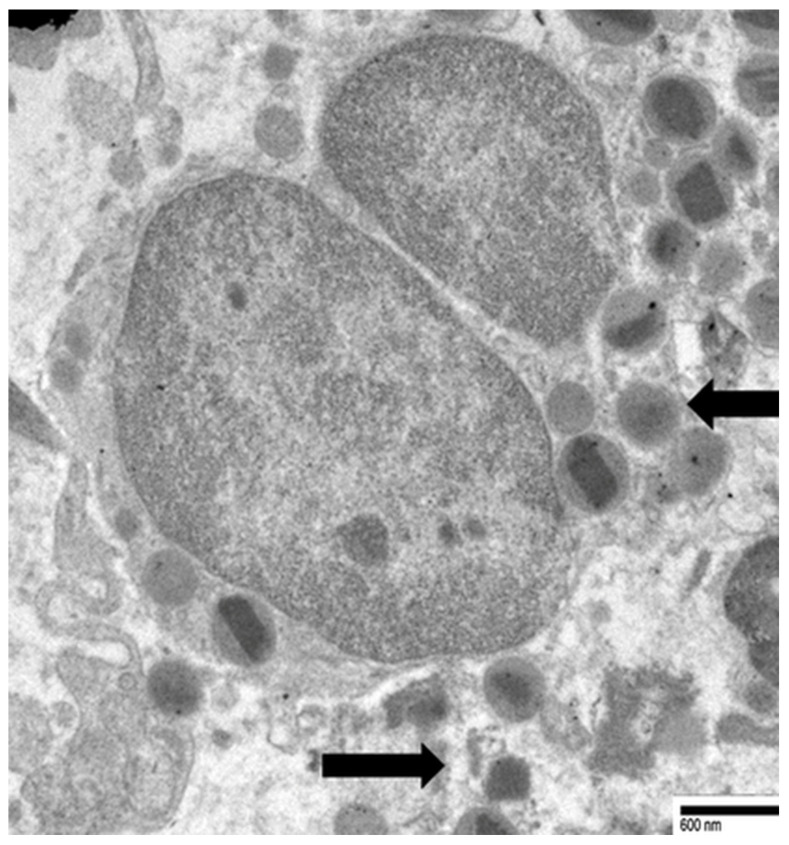
Transmission electron micrograph showing eosinophil cytolytic degranulation: free extracellular granules with intact crystalloid cores, released by cell lysis rather than classical exocytosis.
Caruso, Caruso, Rigoli et al 2025, Cancers · CC BY 4.0 · source
Illustrations
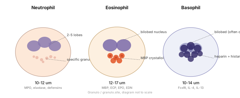
Neutrophil, eosinophil, and basophil side by side, with nuclear shape and granule types labeled.
Granulo / granulo.site · CC0
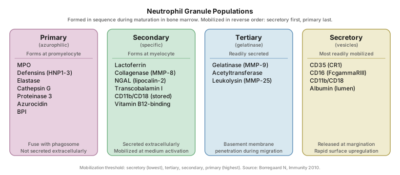
Neutrophil granule populations: azurophilic, specific, gelatinase, and secretory vesicles, with their contents and the order they are released.
Granulo / granulo.site · CC0
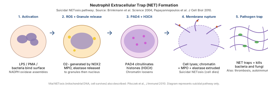
NET formation: activation, oxidant burst, elastase and MPO entering the nucleus, PAD4 histone citrullination, chromatin release, and bacteria trapped.
Granulo / granulo.site · CC0